Close
Close Choose language
Select

Dewax and HIER Buffers from Epredia

Author: Jennifer H. Freeland

When conducting all-in-one deparaffinization and heat-induced epitope retrieval (HIER) procedures for immunohistochemistry (IHC), laboratories can experience inconsistent staining. This typically originates from slides becoming re-coated with molten paraffin when they are removed from the HIER buffer. This causes the IHC reagents from being able to interact with the sample’s antigens. Consistency in IHC staining is important for accurate patient care and efficient laboratory workflow.

Figure 1: Most all-in-one HIER buffers utilize heat almost exclusively as the mechanism of action to remove paraffin from slides. In this case the paraffin simply melts and floats to the surface, where it can re-coat the slides and negatively impact staining results. When using Dewax and HIER Buffers, complete deparaffinization is achieved by dissolving paraffin into solution using a patented chemistry.

By using Dewax and HIER Buffers, you can rely on uniform staining results. Dewax and HIER Buffers L, M, and H have been developed to simultaneously deparaffinize formalin fixed paraffin embedded (FFPE) sections and perform the HIER procedure. Unlike other dewaxing buffers on the market, the Dewax and HIER Buffers have been specially formulated to perform optimally when slides are held vertically in a buffer bath, such as the PT Module1. These buffers have been validated using the UltraVisionTM Quanto HRP DAB detection system by Epredia and with Epredia rabbit and mouse primary antibodies1.

Figure 2: A – Micrograph colon carcinoma, immunostained with p53 antibody, RM-2103-RQ; epitope retrieval was performed using Dewax and HIER Buffer H, TA-999-DHBH. B – Micrograph of squamous cell carcinoma immunostained with Cytokeratin 5/6, MS-1814-RQ; epitope retrieval was performed using Dewax and HIER Buffer H, TA-999-DHBH. C – Micrograph colon carcinoma, immunostained with p53 antibody, RM-2103-RQ; epitope retrieval was performed using a standard Tris-EDTA buffer, HIER Buffer H, pH 9 after routine deparaffinization through xylene. D – Micrograph of squamous cell carcinoma immunostained with Cytokeratin 5/6, MS-1814-RQ; epitope retrieval was performed using a standard Tris-EDTA buffer, HIER Buffer H, pH 9, after routine deparaffinization through xylene.

Due to their unique chemistry, the Dewax and HIER Buffers are patented formulas2,3,4. The dewaxing action of these buffers comes from a water-soluble organic solvent1. Although less than one percent in a working solution, the solvent is strongly capable of dissolving paraffin into an aqueous solution at moderate temperatures (55-95°C)1. At the same time, additives such as polyethylene glycol help stabilize the cells and tissues, as well as flocculate paraffin debris as the buffers cool1.
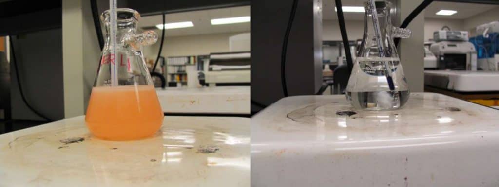
Figure 3: Left – Dewax and HIER L Buffer containing 5 g of molten paraffin at 62oC. The orange solution is homogenous and cloudy, indicating the solubilization of the paraffin. Right – Standard all-in-one citrate epitope retreival buffer containing 5g of molten paraffin at 62°C. The clear reagent shows well-demarcated separation of the molten paraffin at the top with the buffer below.

Product Features and Benefits

  • Easy to use: These buffers are available as 15X concentrates in 100 mL bottles.Each case comes with 10 bottles of 100 mL of buffer. The technologist simply fills the PT Module reagent tank with 1400 mL of distilled or deionized water and adds one bottle of the buffer concentrate to create a working solution.
  • Easy to identify: The buffers are color-coded to assure the technologist that the concentrate has been added to the reagent tank. Since the PT Module carries two tanks, it is possible to simultaneously run two different Dewax and HIER buffers with two different pH ranges. The color coding differentiates the various pH range in each tank.
  • Excellent shelf life and flexible storage: The buffers have a shelf life of 18 months and can be stored at room temperature.
  • Increased throughput: Each of the Dewax and HIER Buffers, once reconstituted, can be reused for up to three PT Module runs of 24 slides each. That is a total throughput of 72 slides per bottle, and 720 slides per case.
  • Verified product performance: Dewax and HIER Buffers have been formulated for optimal performance with eleven (11) different Thermo Scientific paraffin types, see Table 1.
Table 1: Paraffin compatibility chart.

Discover new ways to deliver cleaner slides. Learn more today by contacting your Epredia representative.

Product Descriptions

TA-999-DHBL: Dewax and HIER Buffer L is a low pH (6.0 to 6.4) citrate based buffer. The concentrated solution is a clear, orange liquid.
TA-999-DHBM: Dewax and HIER Buffer M is a medium pH (7.6 to 8.0) EDTA based buffer. The concentrated solution is a clear, purple liquid.
TA-999-DHBH: Dewax and HIER Buffer H is a high pH (8.6 to 9.0) Tris-EDTA based buffer. The concentrated solution is a clear, blue liquid.

References

  1. Freeland JH, et al. Water-soluble organic solvent improves paraffin displacement in all-in-one epitope retrieval buffers. Journal of Histotechnology. 36(2):51-58, 2013
  2. Freeland JH and Harvey MA. Dewaxing buffer containing a water-soluble organic solvent and methods of use thereof. European Patent. EP2807249B1. 2018.
  3. Freeland JH and Harvey MA. Dewaxing buffer containing a water-soluble organic solvent and methods of use thereof. Patent, People’s Republic of China. CN104334717A. 2015.
  4. Freeland JH and Harvey MA. Dewaxing buffer containing a water-soluble organic solvent and methods of use thereof. US Patent. US8753836B2. 2014.
The Future of Pathology is Digital: 3DHISTECH’s Digital Pathology Solutions

Since the turn of the century, the introduction of digital technology into life sciences fields has resulted in improved speed, efficiency, and a reduction in human error.1 Recently, these advancements have been introduced into pathology applications. From advanced imaging to automated microarraying systems, digital pathology has enabled laboratories to achieve increased levels of efficiency, collaboration and throughput. Just as important, these technologies have opened new, unprecedented avenues for analyses.

Whole-slide scanning—currently regarded as one of the most promising avenues for the future of research and medicine, especially for cancer and other complex diseases—offers one of the most dynamic applications of digital technology to the pathology workflow. What were formerly time-consuming manual tasks can now be complete faster and more accurately.1, 2 A high-resolution image (brightfield or fluorescence) is created from conventionally prepared histology glass slides and then can quickly be reviewed and analyzed right at the pathologist’s desktop display.

Advanced digital analytic tools streamline the process of counting and identifying cells and other structures from a sample, reducing processing time and eyestrain alike. The digital workflow also affords an easy method for the reference and comparison of multiple slides. These automated processes are key to reducing inter-operator variability in testing, a major source of errors in pathology and histology laboratories, and prepares the pathology lab for the future of increasing use of artificial intelligence tools in tissue analysis.

Expert Opinion: “…hand-counting of tumor cells took an estimated 100 hours per slide compared to 3 minutes using automated image analysis, demonstrating the efficiencies that targeted image analysis techniques can bring.”1, 2

The impact of digital technologies reaches beyond imaging with systems that automate the process of creating tissue microarrays, creating uniform spreads of tissue for DNA extraction, staining, and other procedures allowing for completion far more quickly and accurately than manual processes.

Perhaps the greatest asset that digital pathology offers is collaboration. Digital pathology information, complete with images, tags, markings and notes, can be stored on shared servers and made available to labs anywhere in the world, often within minutes of digitization. Team analyses and diagnostics, second opinions and student training are easier than ever using digitized samples. Unique labeling systems that travel with these images prevent later misidentification no matter how many pathologists receive them. Laboratory information systems can link all digital pathology images for a specific case together to prevent information loss and facilitate cooperation. Real-time teleconsultation and tumor board reviews for oncology cases are easier than ever once pathology samples are digitized. Training new pathology students is much easier when learning how to read an analyze tissue samples does not involve eyestrain-inducing microscopes and detailed information can follow each slide wherever it goes. With physical slides never needing to leave the lab that created them and labeled high-resolution images available from anywhere, pathologists can focus their time where it matters – studying and understanding the relationship between illness and tissue morphology.

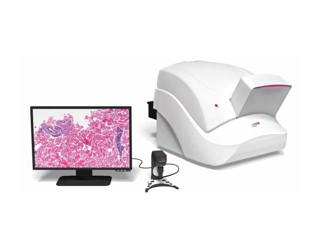
3DHISTECH digital pathology instruments and software, distributed by Thermo Fisher Scientific, features a wide variety of digital pathology tools to improve workflow and bring laboratories into the information age. 3DHISTECH slide scanners include models designed for individual slides, such as the Pannoramic™ Desk II, all the way up to the Pannoramic 1000 that can automatically process up to 200,000 slides per year, and can additionally scan double-width slides commonly used for whole-mount applications.

Most 3DHISTECH slide scanners feature auto-loading cassettes that enable high-throughput processing without direct supervision. For gross examination prior to microscopy, the 3DHISTECH MacroStation is a grossing table with a high-resolution camera that connects to the same computer systems and programs as 3DHISTECH’s slide scanners.

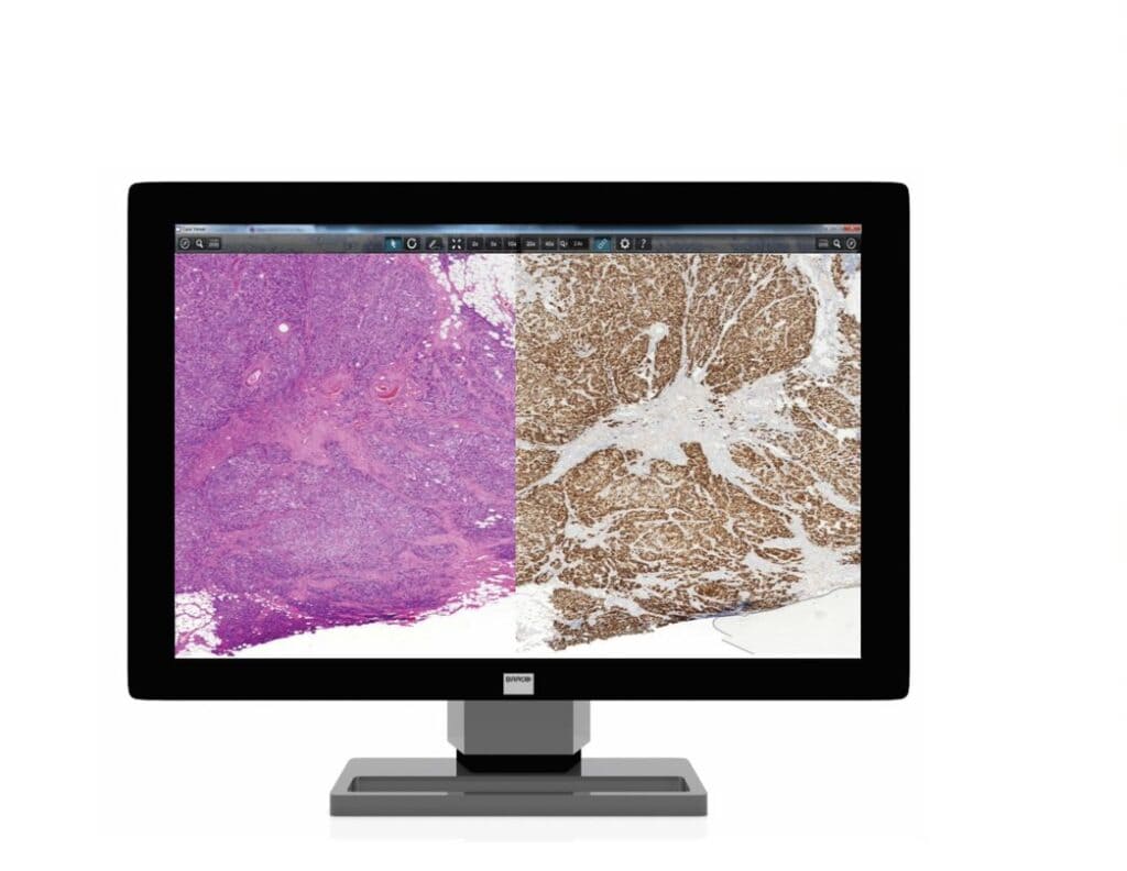
The 3DHISTECH image management system, consisting of the CaseCenter and CaseViewer programs, allows for streamlined and integrated analysis of the images processed by the scanners. CaseCenter links the scanner software to a network and to CaseViewer, and CaseViewer allows the user to review and analyze images stored on the CaseCenter network. CaseViewer allows the user to easily zoom in and out, compare images, add annotations, take measurements, and manipulate the color, contrast, and brightness of the image. QuantCenter, an optional add-on within the CaseViewer software, is designed for quantitative image analysis based on tissue recognition. This software solution can become the center of a digital pathology lab’s operations, and keeps what could otherwise be a bewildering array of samples from multiple sources being treated in multiple ways neat and traceable.

Expert Opinion: “We validated MembraneQuant application of Pannoramic Viewer platform by finding an almost perfect correlation between digital and semi-quantitative evaluation of HER2-IHC slides.3

3DHISTECH also offers the TMA Grand Master and Master II tissue microarrayers. These microarrayers can take hundreds of cores ranging from 0.6 to 2.0 mm in diameter from multiple tissue blocks and create microarrays for future sectioning, staining and analysis. With the same process, they can deposit tissue cores directly into PCR tubes, setting up hundreds of DNA extractions at once. 3DHISTECH’s barcoding system means that slide scanners and other devices can automatically associate each image to match its physical origin, reducing identification errors between users and labs. Furthermore, these images are connected to their case file, enabling entire dossiers to be delivered to collaborators and consultants around the world.

Digital Pathology brings the best of the digital age into the pathology lab. Delivering speed, efficiency, portability, organization, and integration, 3DHISTECH digital pathology instruments and software can take your pathology lab to the next level. Discover ways you can improve your laboratory’s digital pathology or histology workflow, with solutions from 3DHistech, click here.

3DHISTECH instruments are currently available for Research Use Only in the United States.


Contact us for more information

References

1. Griffin, J. & Treanor, D. (2017) “Digital pathology in clinical use: where are we now and what is holding us back?” Histopathology 70 (pp. 134–145).
2. Hamilton PW, Wang Y, Boyd C et al. (2015) Automated tumor analysis for molecular profiling in lung cancer. Oncotarget 6 (pp. 27938–27952).
3. Micsik, T. et al. (2013) “Is HER2 amplification predictable by digital immunohistochemistry?” Diagn. Pathol. 8 (p. S14).

You are leaving the full Epredia.com website. You will be redirected to the [language] site with limited information on our solutions for your laboratory. For more information please fill out the 'contact us' form and a sales representative will answer all of your questions.

Go Back
Continue

您将离开 Epredia.com中文网站,跳转到[language]文网站。网站内的实验室解决方案内容有限。如需了解更多信息,请填写联系表格,我们的销售代表将与您联系, 回答您的所有问题。

后退
继续

Sie verlassen die deutsche Website von Epredia.com. Sie werden auf die [language]-Website mit eingeschränkten Informationen zu unseren Lösungen für Ihr Labor weitergeleitet. Für weitere Informationen füllen Sie bitte das Kontaktformular aus und ein Vertriebsmitarbeiter wird alle Ihre Fragen beantworten.

Zurück
Weiter

Vous quittez le site complet Epredia.com. Vous serez redirigé vers le site [language] avec des informations limitées sur nos solutions pour votre laboratoire. Pour plus d'informations, veuillez remplir le formulaire "nous contacter" et un représentant des ventes répondra à toutes vos questions.

Retourner
Continuer

Stai uscendo dal sito Epredia.com completo. Verrai reindirizzato al sito [language] con informazioni limitate sulle nostre soluzioni per il tuo laboratorio. Per ulteriori informazioni, compila il modulo "contattaci" e un rappresentante commerciale risponderà a tutte le tue domande.

Restituzione
Continua

Está saliendo del sitio completo de Epredia.com. Será redirigido al sitio [language] con información limitada sobre nuestras soluciones para su laboratorio. Para obtener más información, complete el formulario "Contáctenos" y un representante de ventas responderá todas sus preguntas.

Devolver
Continua