Learn about high throughput water immersion technology for cytology samples
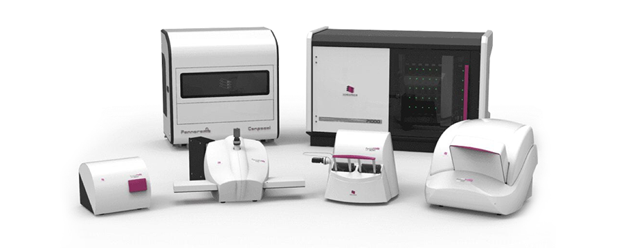
DX Solutions
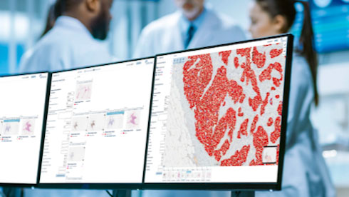
Epredia's comprehensive solution complements your existing workflow, increasing team satisfaction and efficiency.
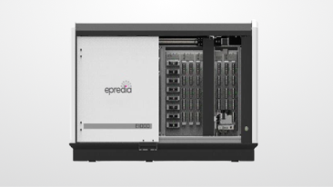
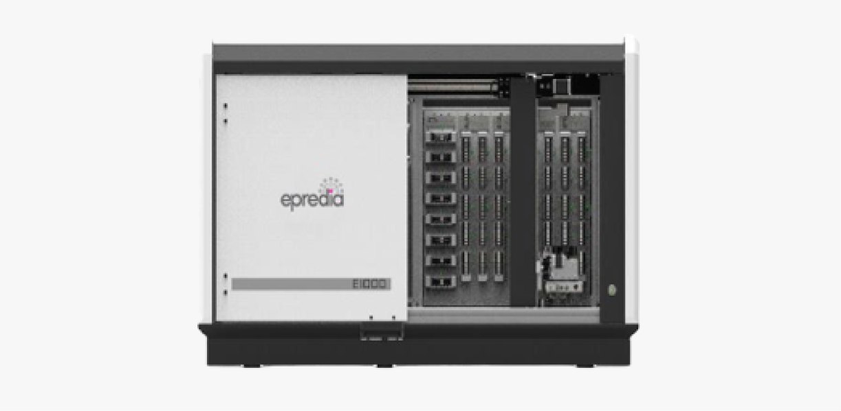
E1000 Dx Solution
BF
Epredia's comprehensive digital pathology solution complements your existing workflow, while increasing your teams satisfaction and efficiency.
More Details
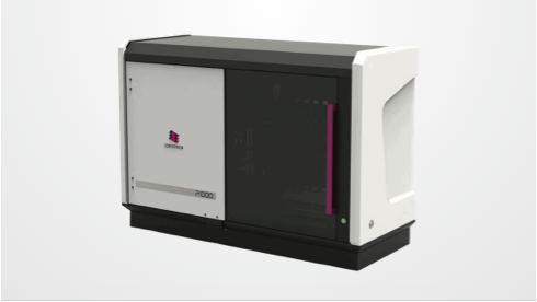
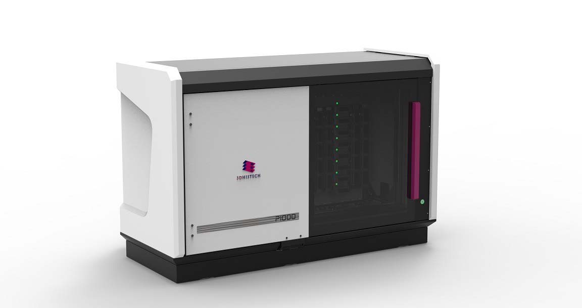
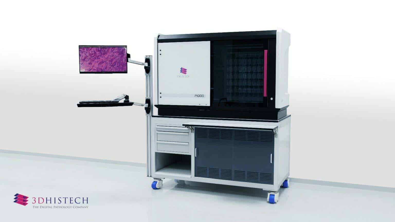
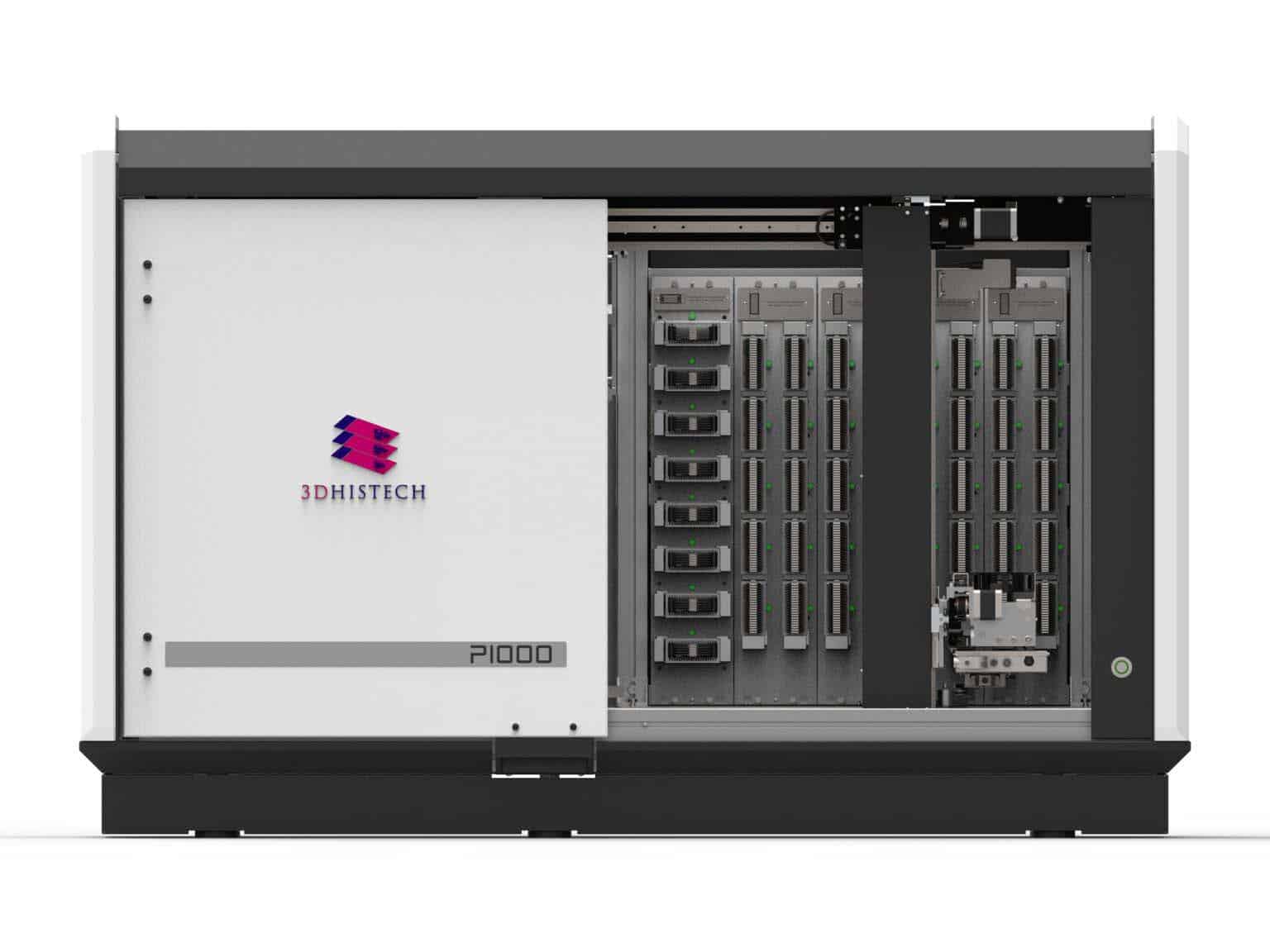
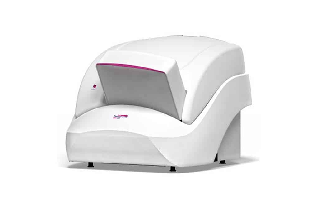
P1000 Dx
BF
1000 slide FLASH scanning for high volume and high throughput labs that don't want to sacrifice flexibility and quality.
More Details
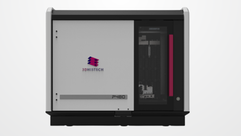
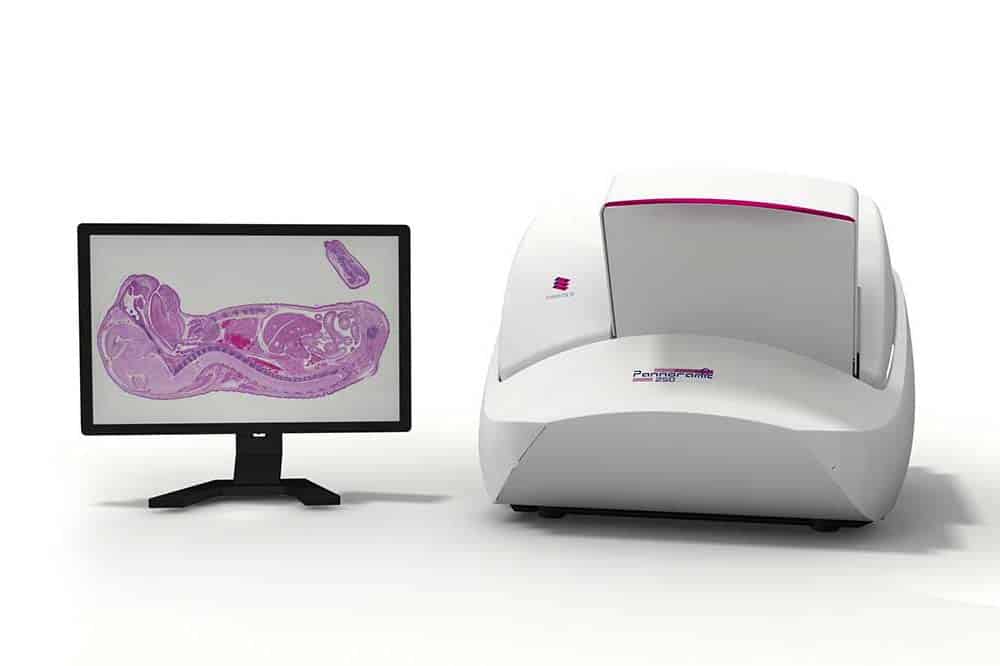
P480 Dx
BF
480 slide scanning for labs that require reliable and automated high through-put while maintaining image quality and flexibility
More Details
P250 Dx
BFFL
For labs that demand BF and/or FL scanning on the same platform with award winning quality and continuous loading of up to 300 slides at FLASH technology speeds.
More Details
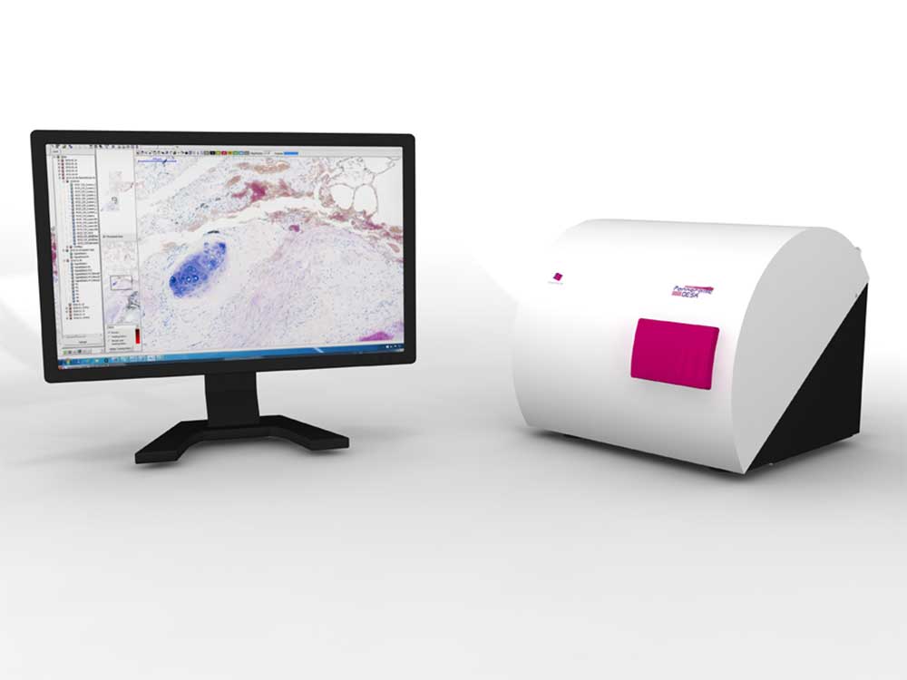
Desk Dx
BF
For labs that need the highest quality imaging capabilities for their remote and frozen section workflow.
More Details
IVD units not for sale in the US, and select other geographies globally. For more information, please contact your local Epredia specialist.
1000 slide FLASH scanning for high volume and high throughput labs that don't want to sacrifice flexibility and quality.
More Details
Pannoramic 480
BF
480 slide scanning for labs that require reliable and automated high through-put while maintaining image quality and flexibility
More Details
Pannoramic 250 Flash III
BFFL
For labs that demand BF and/or FL scanning on the same platform with award winning quality and continuous loading of up to 300 slides at FLASH technology speeds.
More Details
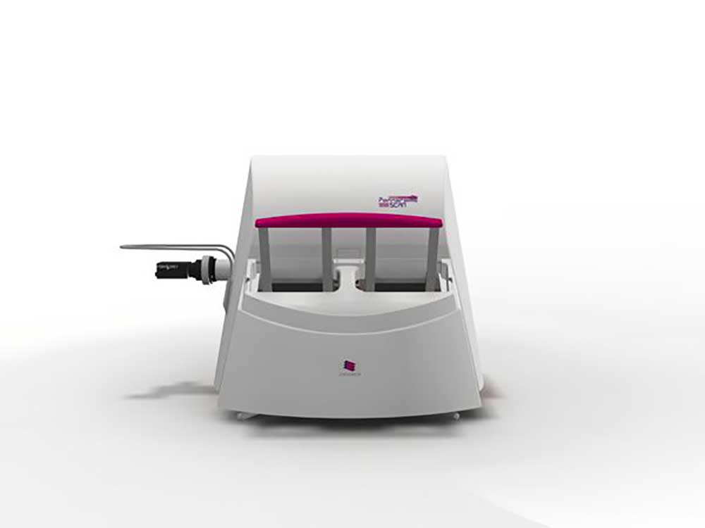
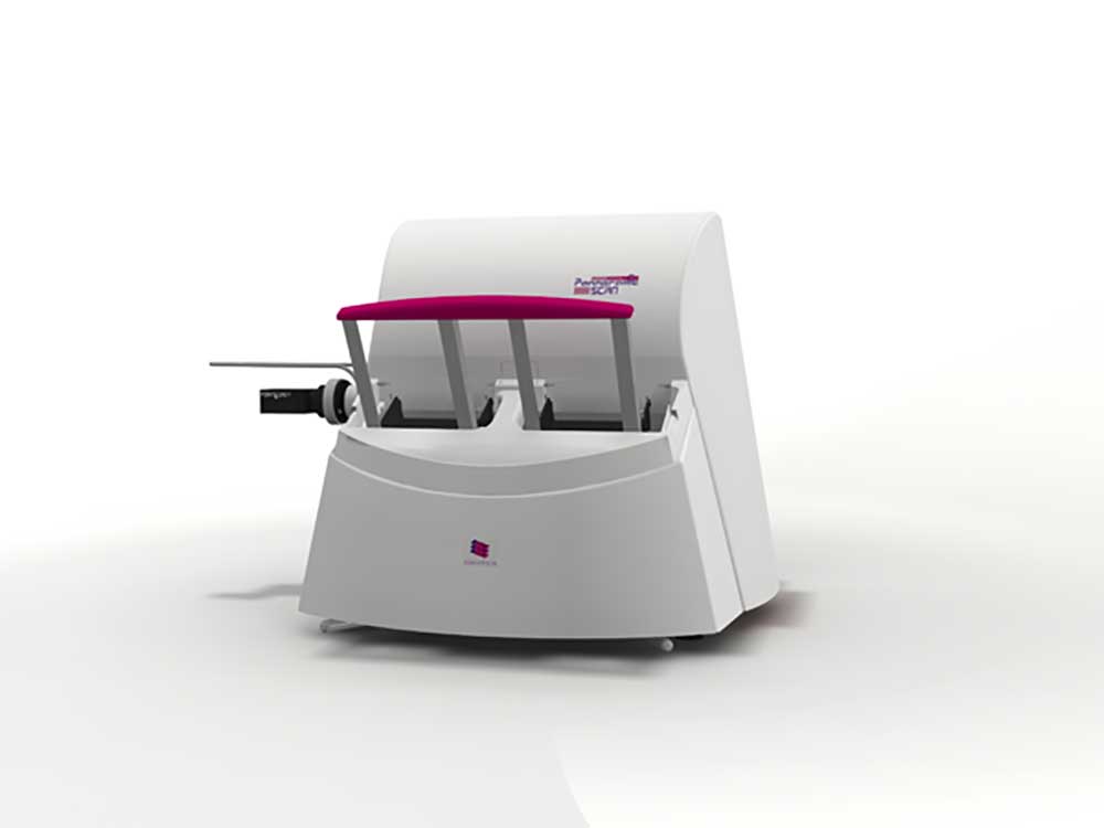
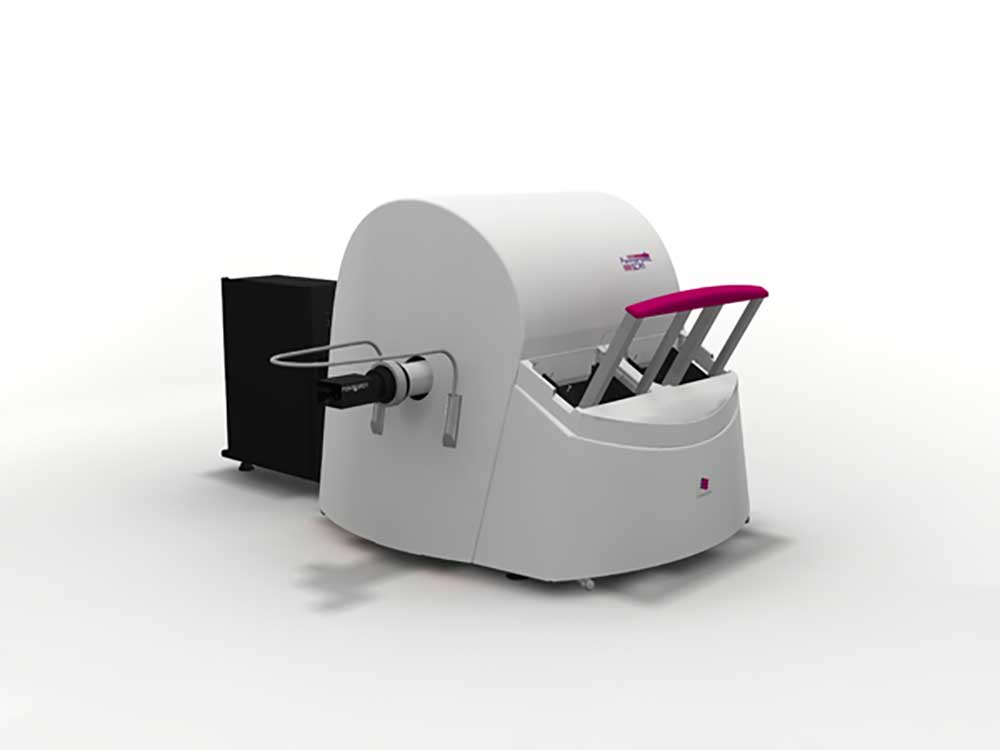
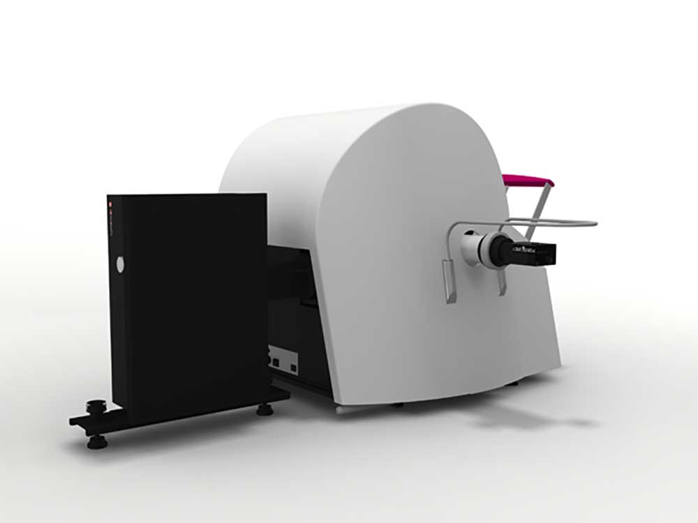
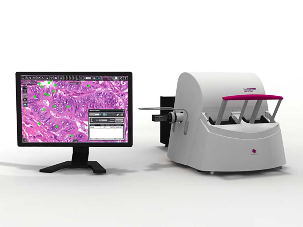
Pannoramic SCAN II
BFFL
For labs that need BF and FL scanning, with continuous workflow loading capabilities of up to 150 slides at a time.
More Details
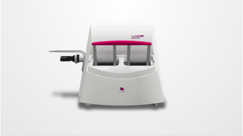
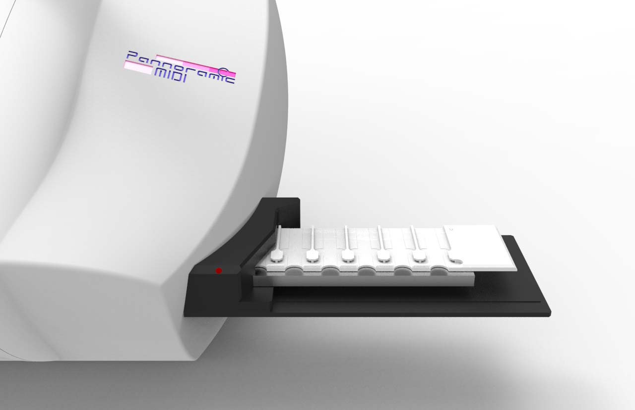
Pannoramic MIDI II
BFFL
For labs that need a compact, flexible scanner with BF and FL scanning capabilities of 12 slide batches.
More Details
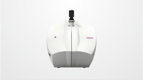
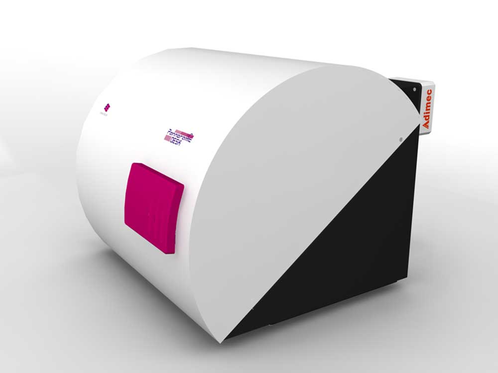
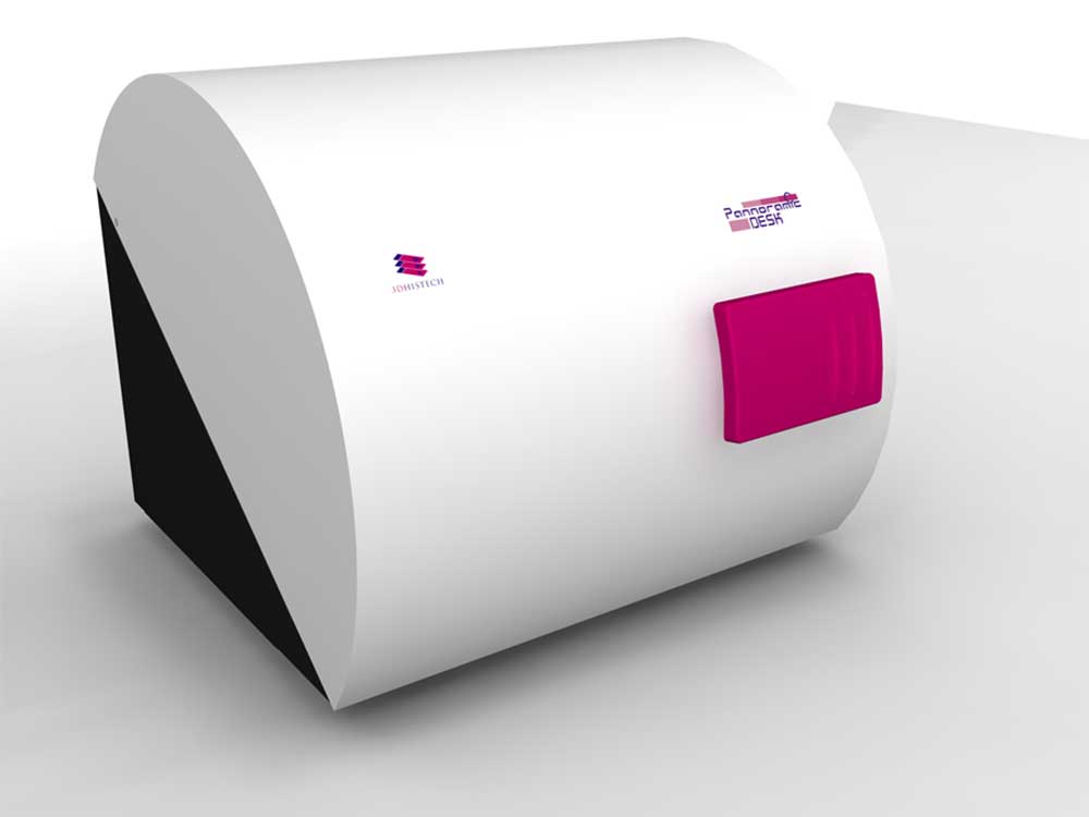
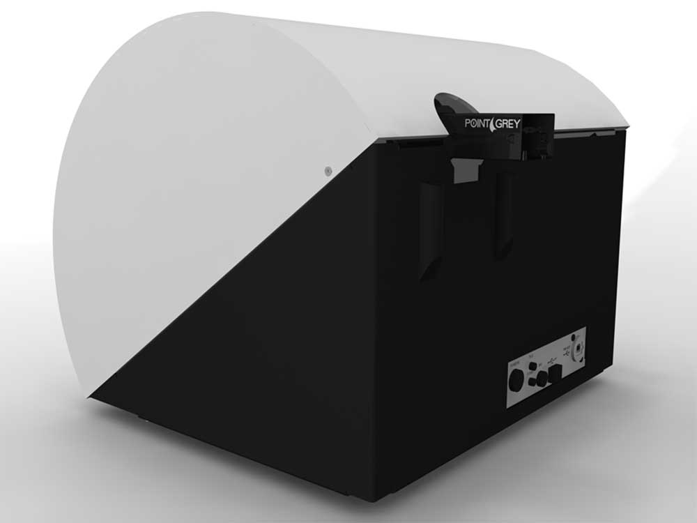
Pannoramic DESK II
BF
For labs that need the highest quality imaging capabilities for their remote and frozen section workflow.
Software solutions provided by Epredia offers pathologists, laboratorians and IT staff with scalable integration and interoperability for an evolving technology landscape.
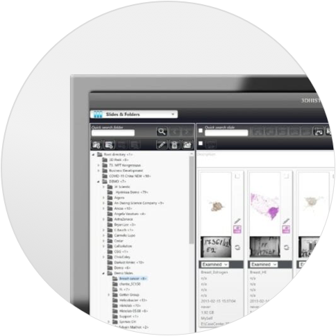
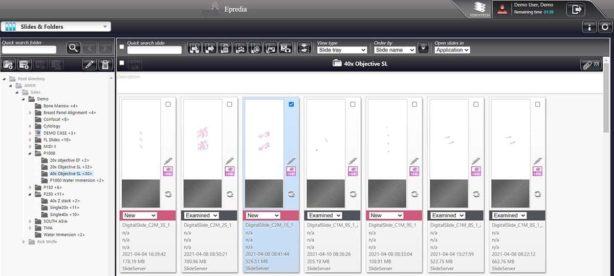
IMS Software
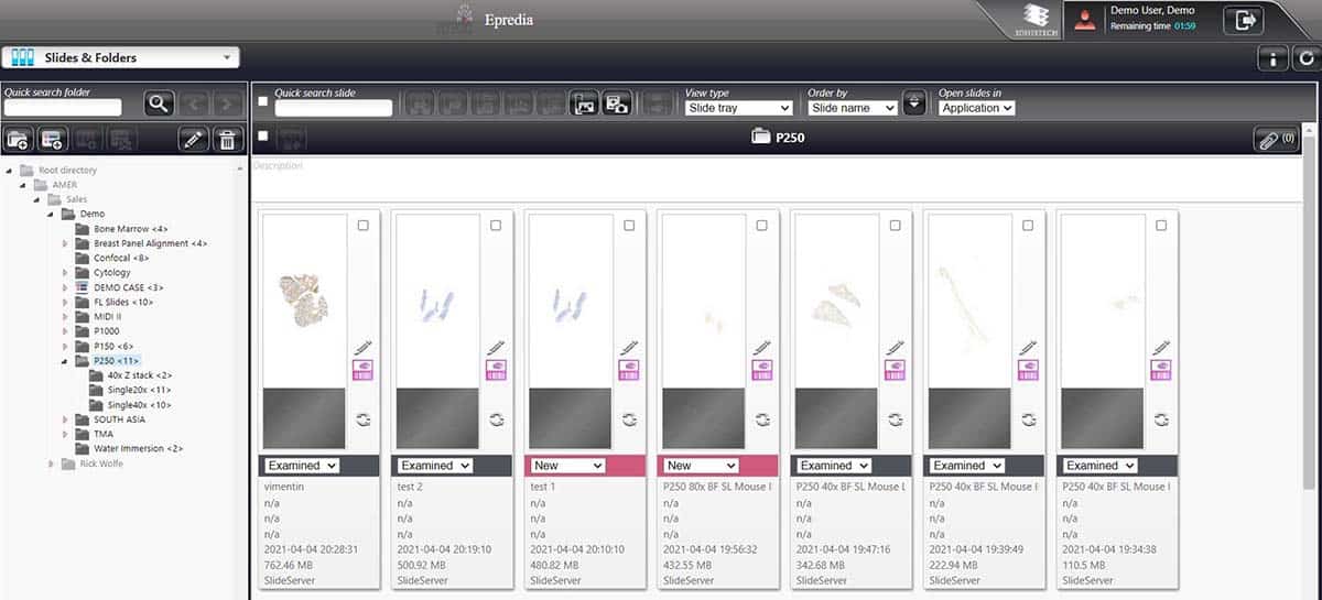
SlideCenter
A powerful, server or cloud based image management system for storing both macroscopic and digital slide images.
More Details
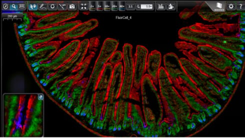
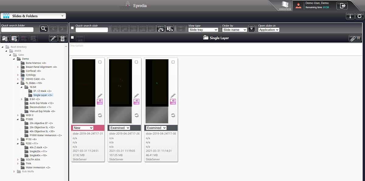
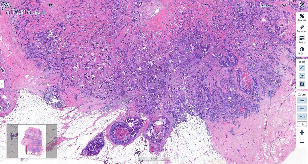
Viewing Software
SlideViewer
Provides researchers and pathologists with a flexible and next generation image viewing experience.
More Details
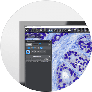
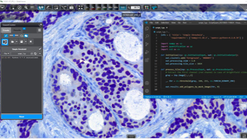
IA Software
QuantCenter
A multi-module image analysis platform designed for image quantification in histopathology, FISH and molecular pathology.
More Details
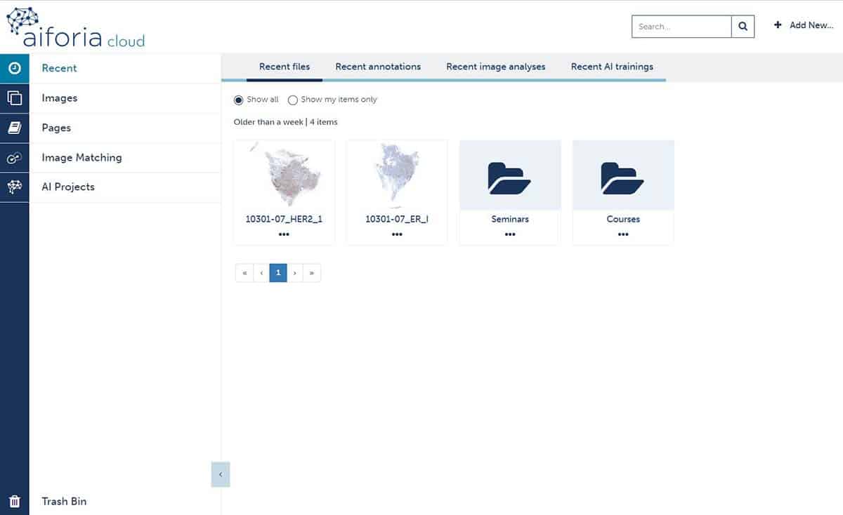
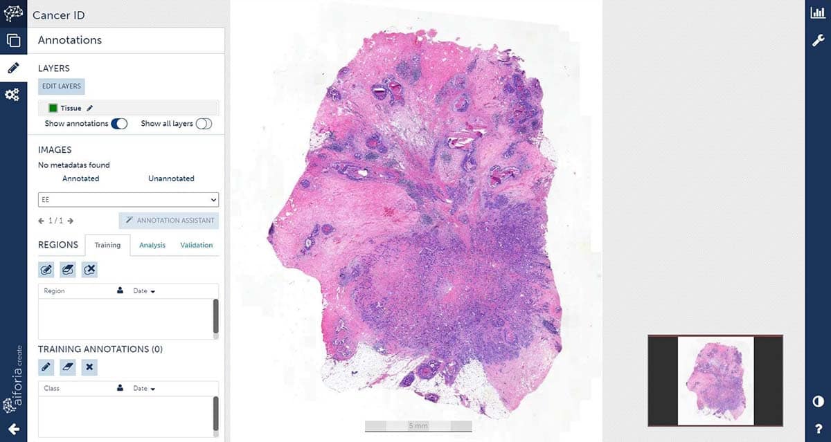
AI Software
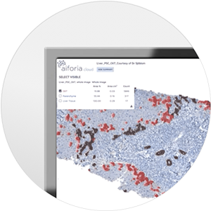
Aiforia
Equips pathologists and scientists in preclinical and clinical labs with powerful deep learning and cloud-based technology to advance their image analysis tasks and workflows.
More Details
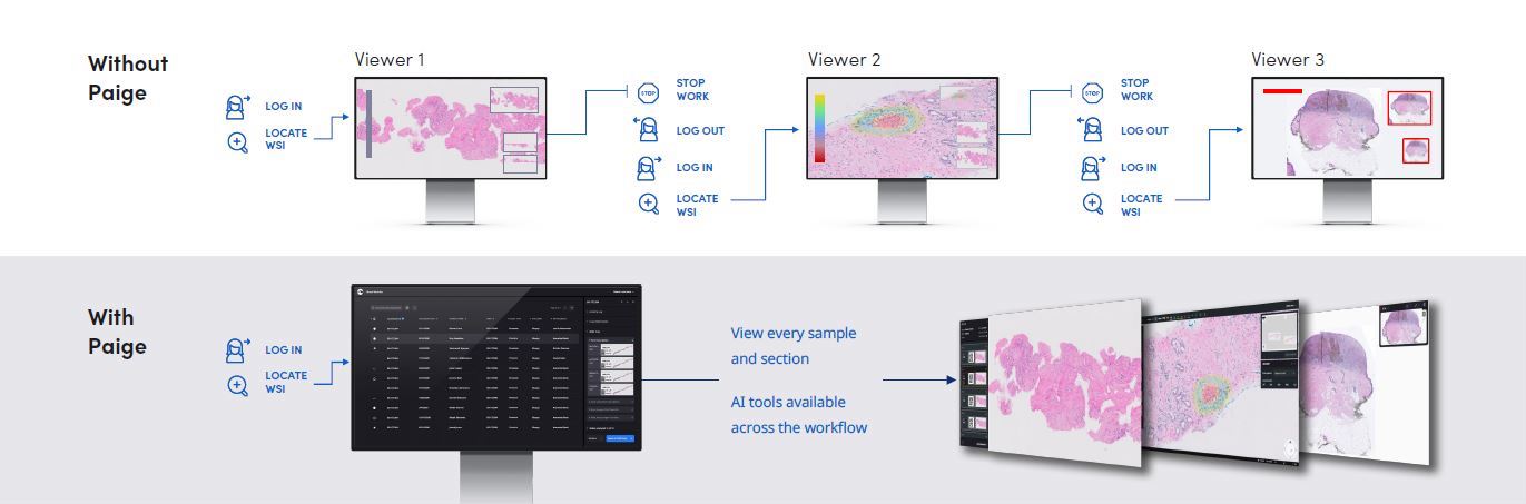
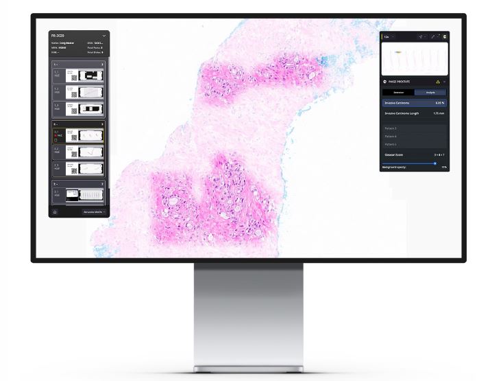
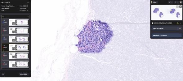
Paige
Provides additional information from digital slides to help pathologists and scientists perform their diagnostic work efficiently and confidently.
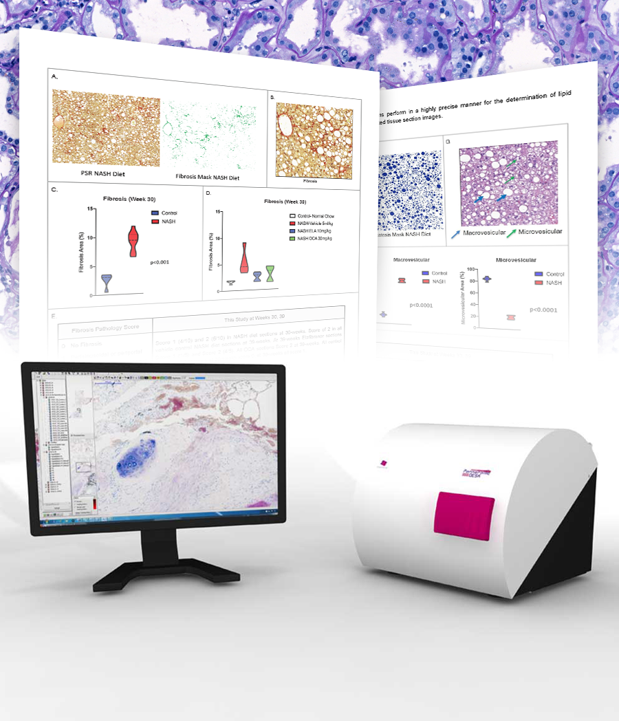
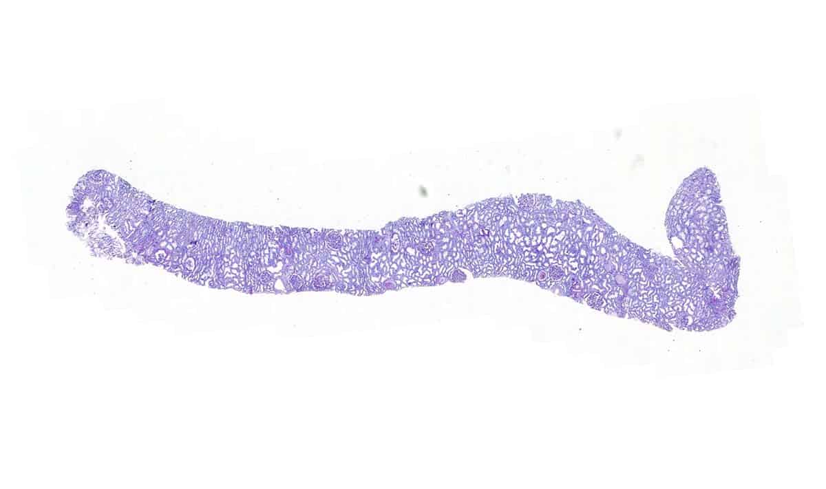
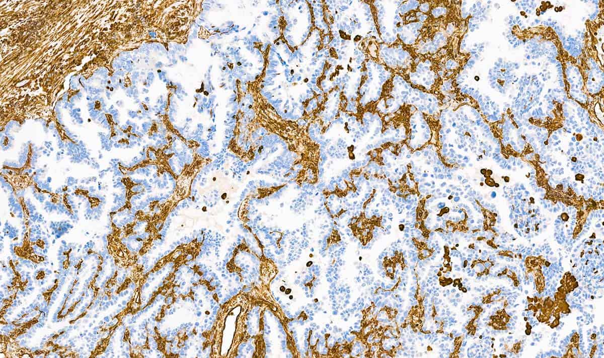
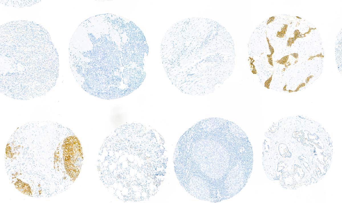
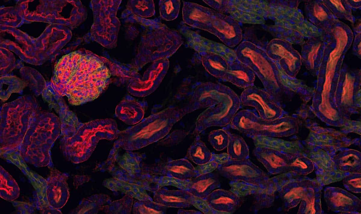
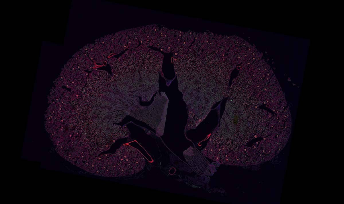
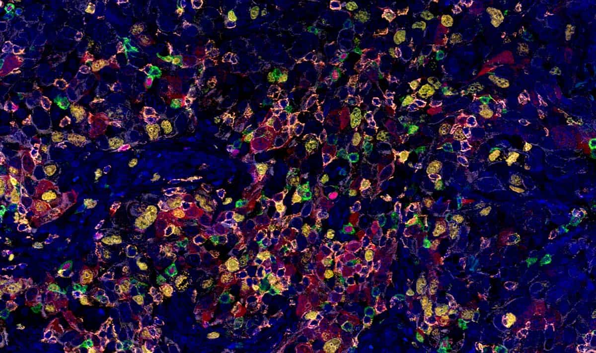
Using Digital Whole Slide Scanning of Liver Tissue and AI-Enabled, Quantitative Histopathological Analysis
The study described in this paper is the result of a unique collaboration between several companies with combined goals of providing the NASH research community with a time-efficient, flexible, off-the-shelf diet-induced mouse NASH B6 Model (Taconic Biosciences, Inc.) that can be utilized in directed research studies for therapeutic drug development (Explora BioLabs).
For research and education use only, not for use in diagnostic procedures in the U.S. This product has not been approved or cleared as a medical device by the U.S. Food and Drug Administration. For Europe this product has been designed for diagnostic purposes, although the medical device registration process is pending in Europe.
Need help finding the right solution for your laboratory?
Contact us.
Thank you for getting in touch
We appreciate you contacting us about digital pathology solutions from 3DHISTECH™ and Epredia™.
Your message has been submitted successfully and one of our representatives will contact you.
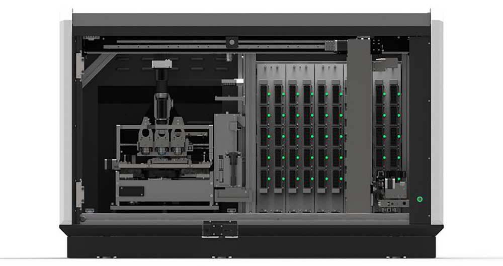
300-slide capacity and continuous loading with vertical slide arrangement.
Award-winning, exceptional image quality for both brightfield and up to nine fluorescent filter positions available for single and multiple band cubes with advanced FISH scanning technique.
Pulsed Xenon FLASH light source for high-speed brightfield scanning.
Up to 90x brightfield and 60x fluorescent magnification by default.
Darkfield preview for easy localization of fluorescent samples.
Brightfield slide scanning in one minute at 40x resolution.
Motorized objective and camera changer.
Automatic slide loading, previewing, barcode reading and scanning.
150-slide capacity and continuous loading with vertical slide arrangement.
Exceptional image quality for both brightfield and up to nine fluorescent filter positions available for single and multiple band cubes with advanced FISH scanning technique.
Up to 90x brightfield and fluorescent magnification by default.
Motorized objective changer.
One high-quality monochrome camera is used for both brightfield and fluorescence with unique three-channel brightfield light source.
Automatic slide loading, previewing, barcode reading and scanning.
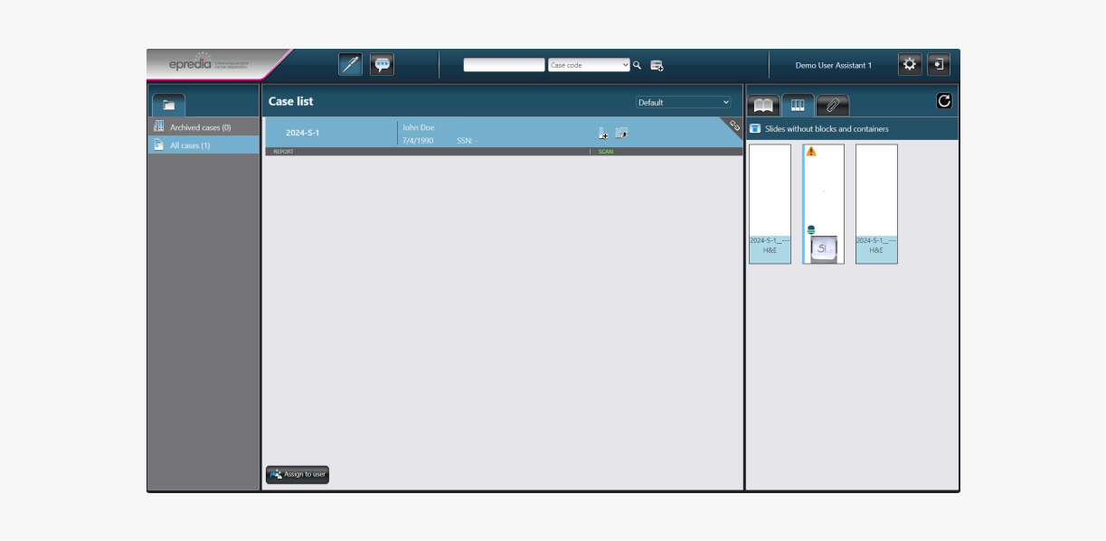
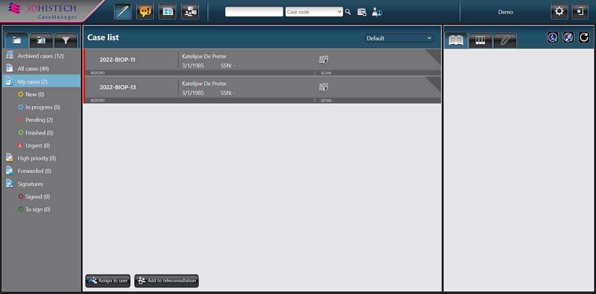
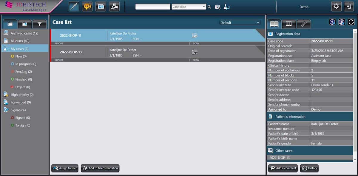
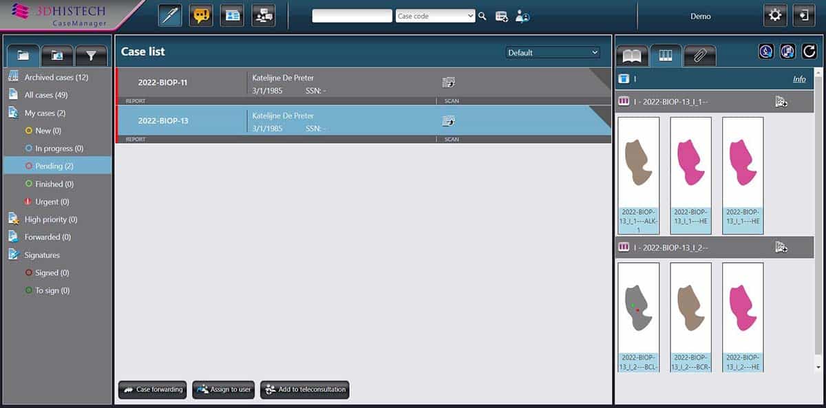
Feature packed. See why our users love CaseManager.
Sending special examination requests to the laboratory information system.
Integration with ClinicalViewer: Patient and staining data from CaseManager is displayed on the top of the viewer’s user interface.
Integration with IVD approved Diagnostic Applications: Results of the automated image analysis are displayed in CaseManager.
Communication with 3DHISTECH’s CentralLogService application to log user and system activity.
Multi-purpose digital pathology workflow support: CaseManager supports the digital pathology workflow with final report in the fields of histopathology, molecular pathology, cytology, and autopsy.
With Focus View, all details of a case are displayed on a single screen. Such details include patient data, additional cases related to the patient, diagnosis codes, digital slides, attachments, and the final report, and more.
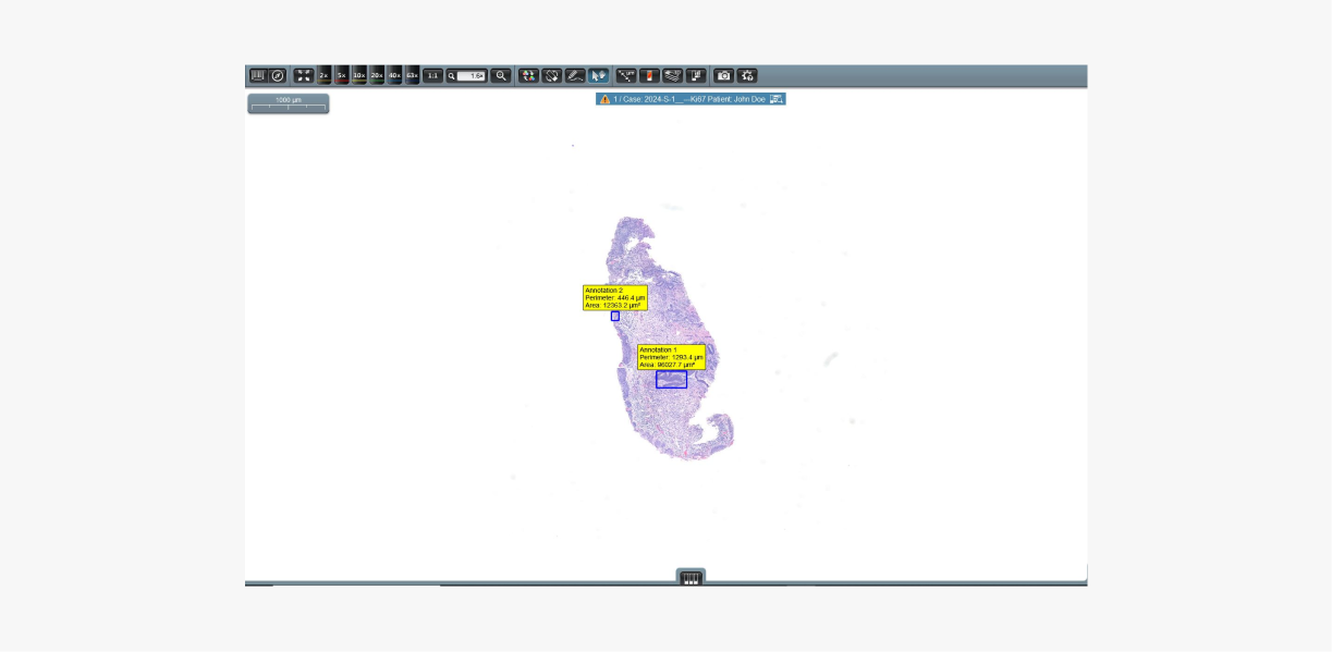
Feature packed. See why our users love QuantCenter.
Molecular pathology
FISHQuant
A powerful cancer and cytogenetic application dedicated to quantify FISH (Fluorescence in Situ Hybridization) signals on tissue samples of solid tumor diseases such as: breast and lung cancer, sarcomas, and lymphomas.
This module is suitable for examination of hematological tumors, FISHQuant classifies the interphase and metaphase cells individually for a comprehensive evaluation.
CISHQuant
Quantify CISH (Chromogenic In Situ Hybridization) stained samples. The algorithm can be calibrated to the stain protocol and quantify by using an integrated color setting tool. This module is suitable for examining gene amplification deletion and chromosome aberration.
The application contains a color adjustment module which can be calibrated to the applied stain protocol and quality.
FISH Quantification
Cancer and cytogenetic application.
FISH quantification on tissue samples of solid tumor diseases, like: breast cancer, lung cancer, sarcoma symptoms, lymphomas.
For hematology type tumors, 3DHISTECH’s FISHQuant application scores the interphase and metaphase cells individually for an even more comprehensive evaluation.
Autofluorescence filtering for FISH (Fluorescence in situ hybridization) samples. As part of QuantCenter, FISHQuant provides a user-friendly, standardized interface and easy navigation bar.
Renewed algorithm for a more sensitive segmentation of nuclei and spots.
Brand new data handling.
Benefit from the fast and safe data processing, easy data visualization and precise data filtering.
IHC Quantification
NuclearQuant
A cell nuclei detection module designed for cell nuclei detection and quantification of IHC stained samples. The algorithm can be calibrated to the stain quality (local laboratory protocol or different stainer) by using an integrated color setting tool.
MembraneQuant
A membrane detection software application can be used for IHC stained histological sample quantification. The algorithm can be calibrated to the stain quality (local laboratory protocol or different stainer) by using an integrated color setting tool.
CellQuant
A cell detection application which is optimal for several IHC quantification.
The application is adequate for cell nuclei, cytoplasmatic and membrane marker quantification. The software reports results based on dedicated scores and positivity ratings of cell nuclei, cytoplasm or membrane signals.
DensitoQuant
An easy to use, fast and accurate, stain-intensity-based IHC quantification tool.
The application identifies the positive stain, based on an automatic color separation method through which individual positive pixels are counted and classified based on intensity and threshold ranges.
The cloud based Aiforia products and services aim to escalate the efficiency and precision of medical image analysis beyond current capabilities, across a variety of fields from oncology to neuroscience and more.
The Paige portfolio includes the Paige Platform, a comprehensive imaging solution comprised of a fast, zero-footprint viewer, storage capabilities and AI-based diagnostic software to help pathologists review cases and support their overall workflow.
5 MP 12-bit camera with RGB illumination (3-chip equivalent)
5 MP 12-bit or 4.2 MP 16-bit camera with RGB illumination (3-chip equivalent)
12 MP 12-bit camera with Xenon Flash illumination
Optical magnification
58x
52x and 110x / 31x and 62x
41x/82x
Highest brighffield scanning speed*
6 min 30 sec
3 min 23 sec
2 min 30 sec
35 Sec (20x) / 1 Min 35 Sec (40X)
30 Sec (20x or 40x)
Average BF file size (native resolution)
2.6 GB (20x) / 7.9 GB (40x)
1.2 GB (20x) / 3.7 GB (40x)
1.25 GB (20x) / 4.5 GB (40x)
Highest throughput/hour
--
15
20
60
72
Fluorescence scanning technology
--
4.2 MP 16-bit camera with wideband / 6-channel LED
Additional 4.2 MP 16-bit camera with 6-channel LED
--
Highest fluorescence scanning speed**
--
6 min @ 31x 22 min @ 62x
5 min @ 31x 15 min @ 62x
--
Dimensions (Wx DxH, cm)
38 x 31 x25
70 x 50 x 50
74 x 53 x 45
68 x 69 x 55
154 x 100 x 91
Weight (kg)
12
23
26
46
270
Technical Specifications
Pannoramic DESK II
Pannoramic MIDI II
Pannoramic SCAN II
Pannoramic 250 FLASH III
Pannoramic 1000
Slide loading capacity
1
12
150 or continuous loading
250 or continuous loading
1000
Double-width slide compatible
Yes
No
No
No
Yes
Objective type
20x (NA 0.8) or 40x (NA 0.95)
20x (NA 0.8) and 40x (NA 0.95)
20x (NA 0.8) and 40x (NA 0.95)
20x (NA 0.8) and 40x (NA 0.95)
20x (NA 0.8) and 40x (NA 0.95)
Brightfield scanning technology
5 MP 12-bit camera with RGB illumination (3-chip equivalent)
5 MP 12-bit or 4.2 MP 16-bit camera with RGB illumination (3-chip equivalent)
5 MP 12-bit or 4.2 MP 16-bit camera with RGB illumination (3-chip equivalent)
12 MP 12-bit camera with Xenon Flash illumination
12 MP 12-bit camera with Xenon Flash illumination
Optical magnification
58x
52x and 110x / 31x and 62x
52x and 110x / 31x and 62x
41x/82x
41x/82x
Highest brighffield scanning speed*
6 min 30 sec
3 min 23 sec
2 min 30 sec
35 Sec (20x) / 1 Min 35 Sec (40X)
30 Sec (20x or 40x)
Average BF file size (native resolution)
2.6 GB (20x) / 7.9 GB (40x)
2.6 GB (20x) / 7.9 GB (40x)
1.2 GB (20x) / 3.7 GB (40x)
1.25 GB (20x) / 4.5 GB (40x)
1.25 GB (20x) / 4.5 GB (40x)
Highest throughput/hour
--
15
20
60
72
Fluorescence scanning technology
--
4.2 MP 16-bit camera with wideband / 6-channel LED
4.2 MP 16-bit camera with wideband / 6-channel LED
Additional 4.2 MP 16-bit camera with 6-channel LED
--
Highest fluorescence scanning speed**
--
6 min @ 31x 22 min @ 62x
6 min @ 31x 22 min @ 62x
5 min @ 31x 15 min @ 62x
--
Dimensions (Wx DxH, cm)
38 x 31 x25
70 x 50 x 50
74 x 53 x 45
68 x 69 x 55
154 x 100 x 91
Weight (kg)
12
23
26
46
270
* 15 x 15 mm area
** 10x10 mm area, 3 filters, 20 ms exposure
×
Disclaimers
FDA Disclaimer
During the COVID-19 pandemic duration, the U.S. FDA guidelines for digital pathology have been amended to allow for digital pathology solutions* in remote settings, that are not currently 510(k) cleared for diagnostics. We are ready to assist you with a special solution that includes a 3DHISTECH Pannoramic 1000 whole slide scanner with CaseCenter and CaseManager software, along with the integration services for a special package price.
Due to the need for compliance with the FDA, this offer is only available to U.S. customers. No modifications to the scanner and software are possible for this remote clinical purpose.
For research use only, not for use in diagnostic procedure.
This product is not available in all regions.
Please reach out to your local sales representative for more information